When it comes to physical activity, shoulder and bicep injuries are some of the most common injuries that occur at the gym. Whether you’re a beginner or an experienced weightlifter, understanding how to identify and treat these injuries is essential for a speedy recovery. In this blog post, we will cover the most common shoulder and bicep injuries that gym-goers experience, and provide advice on how to treat them. Keep reading to learn more about shoulder and bicep injuries, and how to get back in action quickly!
- 1. Introduction to Shoulder and Bicep Injuries in the Gym
- 2. Causes of Shoulder and Bicep Injuries
- 3. Symptoms of Shoulder and Bicep Injuries
- 4. Diagnosis of Shoulder and Bicep Injuries
- 5. Treatment Options for Shoulder and Bicep Injuries
- 6. How to Prevent Shoulder and Bicep Injuries in the Gym
- 7. Advice for Moving Forward After a Shoulder or Bicep Injury
1. Introduction to Shoulder and Bicep Injuries in the Gym
Shoulder and bicep injuries can occur in the gym, but proper diagnosis and treatment can help prevent long-term damage. To diagnose a shoulder or bicep injury, it is important to look for signs of pain, swelling, or difficulty moving the joint. If any of these are present, it is important to seek medical attention as soon as possible. Treatment for shoulder and bicep injuries will depend on the severity of the injury and the type of damage caused. In many cases, rest is the best treatment for a shoulder or bicep injury. This means avoiding any activities that could cause further injury. Ice can be used to reduce pain and swelling, and over-the-counter pain medications can also help. In more severe cases, physical therapy or surgery may be required.
If you experience any pain, swelling, or difficulty moving your shoulder or bicep, it is important to stop exercising immediately and seek medical attention. With proper diagnosis and treatment, shoulder and bicep injuries can usually be prevented from becoming long-term problems.
2. Causes of Shoulder and Bicep Injuries
The most common shoulder and bicep injuries in the gym are caused by overuse or incorrect form while exercising. Overuse injuries occur when too much strain is placed on the muscles, tendons, and ligaments of the shoulder and bicep. This can happen when a person does too many repetitions of an exercise, or does too many sets of exercises in a row. Incorrect form can also lead to shoulder and bicep injuries, as it can put excessive strain on the muscles and tendons, leading to inflammation and pain. Poor posture can also lead to injuries in this area. If the person is not standing up straight and using proper form, the muscles of the shoulder and bicep can be placed under too much strain. Lastly, weight lifting with poor technique can cause injury in the shoulder and bicep area. If the person is lifting too much weight or with incorrect form, they may end up straining or tearing the muscles or tendons of the shoulder and bicep.
3. Symptoms of Shoulder and Bicep Injuries
Shoulder and bicep injuries are common in the gym, but they can be tricky to diagnose. It’s important to pay attention to your body and know what symptoms to look out for so you can get the treatment you need. Common symptoms of shoulder and bicep injuries include pain, swelling, tenderness, and stiffness. You may also have difficulty moving your arm or have a decreased range of motion. You may also experience a popping or snapping sensation when trying to move your arm.
If you are experiencing any of these symptoms, it is important to see a doctor right away. Your doctor will perform a physical exam and may order imaging tests such as an X-ray or MRI to help diagnose the injury. After the diagnosis is made, your doctor can determine the best course of treatment, which may include rest, physical therapy, medications, or surgery.
4. Diagnosis of Shoulder and Bicep Injuries
Diagnosis of shoulder and bicep injuries can be tricky, so it is important to pay attention to any pain or discomfort you may be feeling in these areas. If the pain is severe, or if the range of motion is limited, you should seek medical attention right away. Your doctor will likely do a physical exam and ask questions about your injury, such as when it started and how it has progressed. They may also order imaging tests, such as an X-ray or MRI scan, to get a better look at the affected area.
If the injury is caused by a specific activity, such as weight lifting, your doctor may also recommend that you stop doing the activity until your injury has healed. They may also prescribe physical therapy or suggest other treatments to help reduce your pain and improve your range of motion.
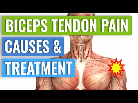
5. Treatment Options for Shoulder and Bicep Injuries
Treatment Options for Shoulder and Bicep Injuries The treatment for shoulder and bicep injuries depends on the severity of the injury. Most minor injuries can be treated with rest, ice, and physical therapy. Rest is important to allow the body to heal and prevent further damage. Ice can help reduce swelling and pain, while physical therapy can help restore movement and strength in the affected area.
If the injury is more serious, other treatments may be necessary. Surgery may be needed to repair damaged tendons or muscles. Medication, such as nonsteroidal anti-inflammatory drugs (NSAIDs), may be prescribed to reduce pain and swelling. Steroid injections can also be used to reduce inflammation and speed up healing.
Rehabilitation is important for any shoulder or bicep injury. A physical therapist or trainer can help create a rehabilitation program tailored to the individual’s needs. This program may include exercises, stretches, and other activities that can help strengthen the muscles and restore normal range of motion. With proper diagnosis, treatment, and rehabilitation, most shoulder and bicep injuries can heal completely. However, it is important to take preventive measures to avoid future injuries. Proper warm-up exercises before any workout and using the correct form for each exercise are key to avoiding injury.
6. How to Prevent Shoulder and Bicep Injuries in the Gym
Shoulder and bicep injuries are common in the gym, but there are ways to prevent them. Before working out, it is important to properly warm up and stretch the muscles. This will help to increase flexibility, which will decrease the risk of injury. It is also important to use proper form when exercising, as this will help reduce strain on the muscles and tendons. Additionally, it is important to not overwork the muscles, as this can lead to fatigue and injury. If a person does experience any pain during or after a workout, it is important to stop the activity and seek medical advice.
7. Advice for Moving Forward After a Shoulder or Bicep Injury
Once you have been diagnosed with a shoulder or bicep injury, it is important to move forward with treatment. Depending on the severity of the injury, your doctor may recommend rest, physical therapy, and/or surgery. Resting your shoulder or bicep is the first step in healing and can help to reduce inflammation. Physical therapy will help to strengthen the muscles and tendons around the injured area and reduce pain. Surgery may be needed if the injury is severe or if there is a tear in the tendon. Once the injury has healed, it is important to take steps to prevent future injuries. Start by increasing strength and flexibility in your shoulder and bicep muscles. This can be done by doing exercises like arm curls, shoulder presses, and lateral raises. It is also important to use proper form when lifting weights to avoid straining your muscles. Additionally, make sure to warm up before any exercise session to help reduce the risk of injury.
References: Diagnosis and Treatment of Common Shoulder Injuries in the Gym
Treatment of Common Biceps Injuries in the Gym